

Seborrheic Keratosis: How to Recognize It and Options After Diagnosis
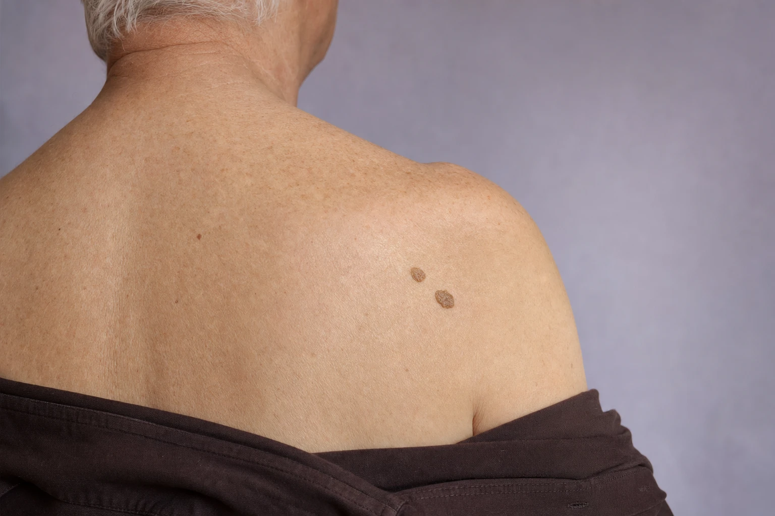
You have noticed a raised, slightly rough brown spot on your back, chest or face that seems stuck on the skin like a drop of wax. You are wondering what it is, whether it can spread or become serious, and whether it is possible to have it removed. This appearance suggests a seborrheic keratosis, a benign growth that appears mostly from middle age onward. However, appearance alone is not enough to identify it: another lesion can look like it, and it takes a medical examination to find out what it is. A seborrheic keratosis is a type of skin growth, but a raised spot is not necessarily a keratosis. CARE MD, in Sainte-Thérèse on Montreal's North Shore, offers a Targeted Lesions consultation for the skin.
- Diagnosis first
- Physician or nurse under supervision
- Sainte-Thérèse
At a glance
Five things to know before deciding- What it is: a benign skin growth, often brown, with a waxy appearance, as if stuck on the skin. It is also called a seborrheic wart, but it is not a wart caused by a virus.
- Contagious or cancerous?: a diagnosed seborrheic keratosis is not passed on to others and does not turn into cancer. However, a very dark keratosis can look like a melanoma: only an examination can show what your spot is.
- Seek care right away if you have a spot that changes, grows quickly, turns black, itches or bleeds, or that looks unusual to you.
- What CARE MD offers: a Targeted Lesions consultation and, after a medical diagnosis and an assessment, removal of seborrheic keratoses, depending on the lesion and the skin.
- Whether to remove it: most keratoses do not need to be removed. Removal can leave the skin lighter, sometimes permanently, and a keratosis can grow back.
What a Seborrheic Keratosis Looks Like
- Chest
- Back
- Abdomen
- Scalp
- Face
- Neck
- Eyelid
A seborrheic keratosis is a benign growth formed by a buildup of skin cells. It is also called a seborrheic wart. It often has a waxy appearance, as if placed on top of the skin, and a rough surface. Its colour ranges from pink through brown to almost black. It can be small and flat at first, then become more raised and wider over the years.
It forms on the chest, back, abdomen, scalp, face, neck or elsewhere on the body, but not on the palms of the hands or the soles of the feet.
You can describe to the doctor the colour of your spot and how raised it is, but these signs are not enough to identify it. The same colour or the same raised appearance can be found in different lesions, and neither a photo nor the mirror is a substitute for an examination.
Why It Appears, and Why You Cannot Catch It
The exact cause of seborrheic keratosis is unknown. It most often forms in middle age or later, and the number of keratoses tends to increase over the years. You may also have a family tendency to develop them; this does not mean you will necessarily get them.
Its other name, seborrheic wart, is misleading. Despite this name, it has nothing to do with the glands that produce sebum, or with warts caused by a virus. It is not contagious: it is not passed on through contact with another person. So seeing new keratoses appear with age does not mean that they have spread.
Exact cause unknown
- Middle age
- Over the years
- Family tendency
It is not contagious
It Does Not Turn Into Cancer, but It Can Look Like It
Once diagnosed, a seborrheic keratosis does not turn into skin cancer. In fact, seborrheic keratoses are much more common than skin cancers. The risk lies in confusing it with other lesions: a very dark keratosis can look like melanoma, a skin cancer. Even if you think you recognize a keratosis, you cannot know whether a spot is benign without having it examined. Have your family doctor or a dermatologist examine a brown or black spot that changes, to check that it really is a seborrheic keratosis.
Another lesion can cause confusion. A dry, flat, rough spot that peels can be an actinic keratosis, some of which develop into skin cancer. Have a doctor examine a dry, rough spot before considering having it removed because of how it looks.
- What you see
-
A raised brown spot, as if stuck on the skin
- What only the doctor can tell on examination
-
- Seborrheic keratosis
- Solar lentigo
- Actinic keratosis
- Sometimes, melanoma
Seborrheic Keratosis or a Lesion to Watch: When to See a Doctor
A brown spot is not always a seborrheic keratosis. An age spot, also called a solar lentigo, is generally a flat brown or black spot that appears on areas of skin exposed to the sun. A seborrheic keratosis, on the other hand, often becomes raised and takes on a waxy appearance. Whether a spot is raised is not enough on its own to distinguish between the two: a keratosis can also be flat at first. Telling them apart requires a medical examination. When a spot thought to be a lentigo looks irregular or unusual, a biopsy, meaning the analysis of a small skin sample, may be needed to confirm the diagnosis and check for melanoma.
Seek care right away
If a spot you thought was a keratosis grows quickly, turns black, itches or bleeds, if it becomes red or painful, or if its appearance changes or seems unusual to you. These signs can occur with skin cancer, but they are not proof of it. Have the spot examined by your family doctor or a dermatologist before considering removal. Make this appointment without waiting for a reply to a message submitted through a form: a written request is no substitute for an examination.
- Grows quickly
- Turns black
- Itches
- Bleeds
- Becomes red or painful
- Changes in appearance
Why Diagnosis Comes Before Any Removal
Before discussing removal, the doctor tries to establish what the lesion is. The doctor examines it, and if any doubt remains about what it is, a biopsy may be done. If the doctor is unsure what the spot is, the diagnosis needs to be clarified first: the spot must not be removed for cosmetic reasons until what it is has been established.
The examination may also result in nothing being removed, or in the decision being postponed. A photo or a message is no substitute for this examination: no diagnosis is made online.
- A biopsy if in doubt
- Nothing removed
- Decision postponed
- No online diagnosis
Why You Should Not Remove It Yourself
Because it seems to be simply sitting on the skin, a seborrheic keratosis sometimes makes you want to scratch it or pull it off. Do not try to remove it yourself: you risk getting an infection. You would also be destroying a spot that has not been examined and that may be something other than a keratosis. If it bothers you, have it examined first; if removal is appropriate, it must then be performed by a professional.
- Infection
- A spot that may be something other than a keratosis
Remove It or Leave It in Place
Not bothersome
A diagnosed seborrheic keratosis is harmless.
Most do not need treatment: you can leave it in place.
Bothersome
Removal can be discussed when it bothers you because it itches, becomes red and tender or catches on clothing.
Appearance
If its appearance is what bothers you, especially on the face, you can also talk to the doctor about it.
However, have any new itching or redness examined before deciding on removal: the doctor must first identify the spot.
Removal at CARE MD
CARE MD offers a Targeted Lesions consultation for the skin. During this consultation, you can talk about the spot that bothers you and your expectations. After a diagnosis and an assessment, CARE MD removes seborrheic keratoses using plasma, cryotherapy or CO2 laser, depending on the lesion and the skin. Meet our professionals, who make up the CARE MD team.
- Prior diagnosis
-
No removal without a diagnosis made by a doctor.
- Who performs the removal
-
A physician or a nurse under medical supervision.
- Scope
-
The skin, including the face.
- Before choosing
-
Have the spot identified, then discuss whether removal is appropriate and the marks it can leave.
- Suspicious lesion
-
Diagnosis comes before any cosmetic request.
- At CARE MD Plasma
-
Ask the doctor about the after-effects and risks
- At CARE MD Cryotherapy
-
Freezing destroys the lesion; a blister, then a scab, can form.
- At CARE MD CO2 laser
-
Ask the doctor about the after-effects and risks
- Described in medicine Electrocautery
-
This is not the same procedure as applying cold.
For a diagnosed keratosis, cryotherapy, one of CARE MD's removal techniques, involves applying liquid nitrogen, a very cold liquid, with a cotton swab or a spray device.
In general, another removal method is electrocautery: after numbing the area with a local anesthetic, the doctor uses an electric current to destroy the lesion.
These removal methods are not a recommendation for your spot. Before choosing, discuss with the doctor what is appropriate for your skin, the possible marks, the number of sessions being considered and how long your skin may take to heal. For plasma or CO2 laser, ask the doctor about the after-effects and risks specific to the technique being considered.
Risks of Removal and What to Expect Afterwards
After removal
- Lighter skin
- Grows back in the same place
- New keratoses elsewhere
After a seborrheic keratosis is removed, the skin can stay lighter than the surrounding skin.
This difference usually fades over time, but it is sometimes permanent. A treated keratosis can also grow back in the same place, and others can continue to appear elsewhere on the body. Removal therefore guarantees neither skin without marks nor an end to keratoses.
After cryotherapy
- Redness
- Swell
- Blister
- Pain
- Scab
- Lighter mark
- Rarely a scar
With cryotherapy, the skin reacts as it would after a burn: it can become red, swell or form a blister.
The cold can be painful during the treatment, and then for a variable length of time. A scab then forms and falls off on its own; how long this takes depends on the area treated. Do not touch it: pulling off the scab makes a scar more likely. The skin can be left with a lighter mark. A scar rarely forms after cryotherapy; it can be white and flat, whatever the colour of the skin.
Infection
- Pain that increases
- Pus
Infection is uncommon after cryotherapy. If the pain increases or you see pus, contact the professional who treated you or your doctor.
For any other technique being considered, ask for an explanation of its specific risks and after-effects before deciding: the reactions described after cryotherapy do not apply to all treatments.
A Seborrheic Keratosis on the Face
The face is one of the places where a seborrheic keratosis can appear, and it is often where its appearance is most bothersome. As elsewhere on the body, before considering removal, have a spot on the face examined if it changes, darkens, itches or bleeds.
If you want a keratosis on your face removed because of how it looks, also weigh up the marks removal can leave. The skin can stay lighter where it was treated, sometimes permanently. After cryotherapy, it can also become darker, especially on brown or Black skin; a white, flat scar rarely forms. Discuss this before choosing a technique; not removing anything is also an option.
For a seborrheic keratosis of the eyelid, a diagnosis is still needed before any removal, even if it looks familiar to you.
- Skin colour after cryotherapy
- Light skinBlack or brown skin
The treated skin can then become lighter or darker, especially on brown or Black skin; this change often improves over time, but it can remain.
Ask Us About Your Seborrheic Keratosis
Tell us where the spot is, when you first noticed it, whether it has changed and whether it itches, bleeds or catches on things. This information helps prepare for your Targeted Lesions consultation. It is not enough to make a diagnosis remotely, and submitting it does not book a removal. The form is not for emergencies: if a spot changes, grows quickly or bleeds, see a doctor without waiting for our reply.
Frequently Asked Questions
Is a seborrheic keratosis contagious?
No. A seborrheic keratosis is not contagious: it is not passed on through contact with another person. The name seborrheic wart is misleading: it is not a wart caused by a virus. If new keratoses appear, it is because their number tends to increase with age, not because they have spread. A medical examination is still needed to confirm that your spot really is a seborrheic keratosis.
Sources2 references
Can a seborrheic keratosis turn into cancer?
A diagnosed seborrheic keratosis does not turn into skin cancer. The danger comes from confusing it with other lesions: a very dark keratosis can look like a melanoma, and a dry, flat, rough spot that peels can be an actinic keratosis, some of which develop into cancer. A spot that changes, grows quickly, turns black, itches or bleeds must therefore be examined by a doctor before any removal, even if you think you recognize it.
Sources2 references
How can I tell whether my spot is a seborrheic keratosis?
Through a medical examination. A seborrheic keratosis often looks waxy and rough, as if stuck on the skin, and its colour ranges from pink to almost black. A solar lentigo is more typically a flat spot on an area exposed to the sun, but a keratosis can also be flat when it first appears. These reference points are therefore not enough: the doctor examines the spot and, if in doubt, may do a biopsy, meaning the analysis of a small skin sample.
Sources3 references
Can you remove a seborrheic keratosis yourself?
No. Scratching, pulling off or trying to remove a keratosis yourself puts you at risk of infection. You would also be destroying a spot that has not been examined, when a lesion that looks like a keratosis can be something else, sometimes a melanoma. Have the spot examined first; if it bothers you, removal should then be discussed after diagnosis. It must be performed by a professional.
Sources3 references
How is a seborrheic keratosis removed at CARE MD?
The first step is to have the spot identified by a doctor. Once a diagnosis has been made, CARE MD removes the keratosis using plasma, cryotherapy or CO2 laser, depending on the lesion and the skin; a physician or a nurse under medical supervision performs the removal. Discuss with the doctor which technique is suited to your skin, the possible marks, the number of sessions being considered and how long healing is expected to take. For plasma and CO2 laser, ask the doctor about the after-effects and risks specific to the procedure. Cryotherapy destroys the lesion by freezing it with liquid nitrogen. In general, a keratosis can also be removed by electrocautery: an electric current destroys the lesion after a local anesthetic. The risks and the care required differ depending on the method and need to be explained for the technique chosen.
Sources1 reference
What are the risks after a seborrheic keratosis is removed?
The skin can stay lighter where it was treated; the difference usually fades, but sometimes remains. A treated keratosis can grow back, and others can appear elsewhere. After cryotherapy, redness, swelling, a blister, pain and then a scab are possible, and pulling off the scab makes a scar more likely. After cryotherapy, the skin can also become lighter or darker, especially on brown or Black skin; this change can be permanent. A white, flat scar rarely forms after this procedure. If the pain increases or you see pus, contact the professional who treated you or your doctor.
Sources3 references
Can a seborrheic keratosis on the face be removed?
Removal on the face can be discussed once a diagnosis has been made. If its appearance bothers you, also consider the possible marks: the skin can stay lighter after removal, sometimes permanently. After cryotherapy, it can become lighter or darker, especially on brown or Black skin; a scar rarely forms and can be white and flat. A spot on the face that changes, itches or bleeds needs to be examined first. A seborrheic keratosis can also be found on the eyelid: CARE MD assesses and treats it after diagnosis, as it does elsewhere on the skin.
Sources5 references
Sources8 references+
- Seborrhoeic keratosis — British Association of Dermatologists (2022) (opens in a new tab)
- Seborrheic keratoses: Signs and symptoms — American Academy of Dermatology (opens in a new tab)
- Seborrheic keratoses: Who gets and causes — American Academy of Dermatology (opens in a new tab)
- Seborrheic keratoses: Tips for managing — American Academy of Dermatology (opens in a new tab)
- Seborrheic keratoses: Diagnosis and treatment — American Academy of Dermatology (2023) (opens in a new tab)
- Liver spots — MedlinePlus, National Library of Medicine (2024) (opens in a new tab)
- Cryotherapy — British Association of Dermatologists (2022) (opens in a new tab)
- How to select a dermatologist — American Academy of Dermatology (2022) (opens in a new tab)
Seborrheic Keratosis: Diagnosis Before Any Removal
Before a decision is made about removal, a doctor must identify the spot. A photo is not enough to make this diagnosis.
Limitations and precautions
Before any treatment, show a doctor a spot that changes, turns black, grows quickly, itches or bleeds.
Do not scratch, pull off or try to remove a keratosis at home.
After cryotherapy, let the scab fall off on its own; if the pain increases or there is pus, call your healthcare provider.
Mention your medications, including non-prescription ones, your vitamins, your supplements and any skin cancers among your close relatives.
The form is not for emergencies.
Neither a waxy appearance, a raised surface nor colour is enough to confirm a seborrheic keratosis; a very dark keratosis and a melanoma sometimes look alike.
Removal can leave the skin lighter, sometimes for a long time. After cryotherapy, it can also become darker or, rarely, be left with a scar. A keratosis can grow back, and others can appear elsewhere.
Choosing a technique requires a discussion with the doctor about your skin and the risks specific to the procedure; the risks of cryotherapy do not apply to all methods.
A medical diagnosis is still needed before any removal.