


Cherry Angioma (Ruby Spot): How to Recognize It and Options After Diagnosis
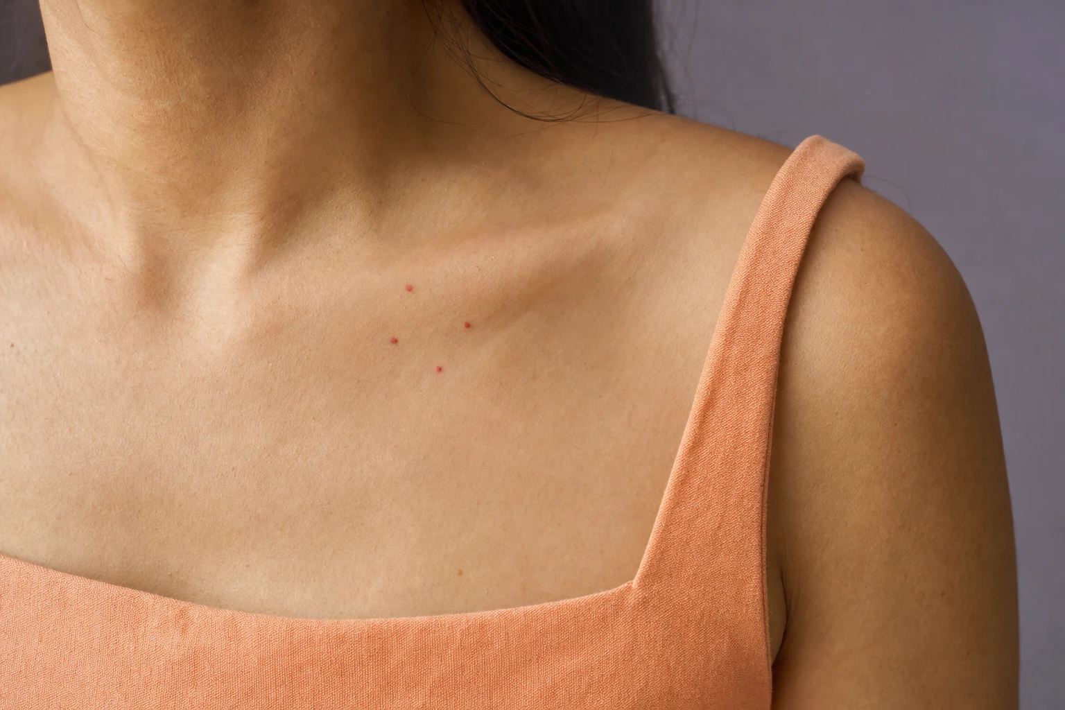
While getting changed, you notice a few small bright red spots on your chest, abdomen or arms, sometimes slightly raised. Some have been there for years, others seem new, and one of them may have bled when a strap or a fingernail caught it. You are wondering what these small red spots are, whether they are dangerous and whether you can have them removed. A cherry angioma is a small benign growth made up of blood vessels. Its appearance helps identify it, but is not enough: it takes a medical examination to confirm what it is. CARE MD, in Sainte-Thérèse on Montreal's North Shore, offers a Targeted Lesions consultation for the skin. The doctor first establishes what the red spot is, before removal is discussed.
- Diagnosis first
- Physician or nurse under supervision
- Sainte-Thérèse
At a glance
Five things to know before deciding- What it is: a cherry angioma, or ruby spot, is a small benign growth made up of blood vessels, usually cherry red, that most often appears on the trunk.
- Why they appear: their cause is unknown; they are more common after age 30 and tend to be inherited.
- Bleeding: a cherry angioma can bleed if it is injured. A red spot that bleeds, changes, grows or itches needs to be examined by a doctor before any removal.
- What CARE MD offers: a Targeted Lesions consultation for the skin and, after a diagnosis and an assessment, removal of a cherry angioma, depending on the lesion and the skin; no red spot is treated automatically.
- Whether to remove it: a cherry angioma generally does not need to be treated. Removal can leave a mark or a scar, and no technique guarantees the result.
What Is a Cherry Angioma (Ruby Spot)?
A cherry angioma, also called a ruby spot, is not cancerous. Cherry angiomas usually have the following characteristics:
- Colour
-
Bright cherry red.
- Surface
-
Smooth or raised.
- Size
-
Small, from the size of a pinhead to a little over half a centimetre.
- Location
-
Almost anywhere on the body, but mostly on the trunk.
These characteristics describe cherry angiomas; they cannot be used to confirm that your red spots are cherry angiomas. A lesion's small size, in particular, is not reassuring on its own: a melanoma can also be small when it is diagnosed. A photo is not enough, and what a red spot is can only be confirmed through an examination.
- What you see
-
A few small bright red spots, sometimes slightly raised
- What only the doctor can tell on examination
-
- Cherry angioma
- Spider angioma
- Broken capillaries
- A melanoma can also be small
Why Cherry Angiomas Appear
- Chest
- Abdomen
- Arms
- Eyelid
Cherry angiomas are more common after age 30. Their cause is unknown, but they tend to be inherited.
Cause unknown
- After age 30
- Inherited
However, a new spot that does not look like the others needs to be examined, even if you already have several.
Are Cherry Angiomas Dangerous, and Why Do They Bleed?
An identified cherry angioma is benign and generally does not need to be treated.
However, it can bleed if it is injured, for example if it gets caught on clothing or nicked by a razor. Even if it was caught on something, show the bleeding spot to a doctor, especially if the bleeding happens again, occurs without an injury or comes with a change in appearance. Once a diagnosis has been made, frequent bleeding can also be a reason to discuss removal.
When to Have a Red Spot Examined Right Away
Seek care right away
If a red spot is new and does not look like the others, if it changes in size, shape or colour, or if it grows, itches or bleeds. These signs are not proof of cancer, but a medical examination is needed before any request for cosmetic removal. Make an appointment with a doctor rather than waiting for a reply to a message submitted through a form: a message is no substitute for an examination. A cherry angioma that has already been identified and stays the same is not one of these signs.
- New, different from the others
- Changes in size, shape or colour
- Grows
- Itches
- Bleeds
Telling Cherry Angiomas Apart From Other Spots and Redness
Other red marks can be mistaken for a cherry angioma.
These differences help describe what you see; they are not enough to make a diagnosis.
Treatment for redness must not be started on a lesion that has not been examined, and a red spot that changes needs to be examined before any decision.
Why Diagnosis Comes Before Any Removal
To identify a cherry angioma, the doctor examines the lesion on the skin; other tests are generally not needed. Sometimes a biopsy, meaning the analysis of a small skin sample, is used to confirm the diagnosis. A cherry angioma is one of the skin growths that CARE MD assesses, all according to the same rule: the doctor first establishes what the lesion is, and removal is discussed afterwards. A spot that raises concern is managed medically, not cosmetically.
The assessment may also show that it is better to leave the angioma in place, or to postpone the decision. Do not try to remove it yourself: a red spot that has not been examined may be something other than a cherry angioma. No diagnosis is made online.
- Sometimes a biopsy
- Do not remove it yourself
- No online diagnosis
- Managed medically, not cosmetically
Cherry Angioma Removal: When to Consider It and Which Techniques Can Be Used
Leave it in place
A cherry angioma generally does not need to be treated, and leaving it in place is a perfectly valid choice.
Discuss removal
Once a diagnosis has been made, removal can be discussed if its appearance bothers you or if it bleeds often.
Suspicious lesion
It is assessed medically, with no cosmetic removal.
In a Targeted Lesions consultation, CARE MD sees people who have this type of red spot. During the consultation, the doctor focuses first on examining the red spot and establishing a diagnosis. After a diagnosis and an assessment, CARE MD removes cherry angiomas using plasma, cryotherapy or CO2 laser, depending on the lesion and the skin. A physician or a nurse under medical supervision performs the removal.
- Prior diagnosis
-
No removal without a diagnosis made by a doctor.
- Scope
-
The skin.
- Consultation
-
Targeted Lesions, to have the red spot examined.
- Choice of treatment
-
It is discussed with you.
- At CARE MD Plasma
-
Ask the doctor to explain its risks and after-effects
- At CARE MD Cryotherapy
-
Cryotherapy is one of the options for removing a cherry angioma.
- At CARE MD CO2 laser
-
Ask the doctor about the specific risks and after-effects
- Described in medicine Vascular laser
-
Among the general options, a vascular laser targets the blood vessels in a lesion.
Cryotherapy
It removes superficial skin lesions by freezing them, most often with liquid nitrogen.
Vascular laser
With a suitable wavelength, meaning light chosen for this target, it damages the vessel walls, which the body then reabsorbs.
This mechanism describes a vascular laser; the laser CARE MD uses to remove cherry angiomas is a CO2 laser.
The choice of device
The choice of device matters, because an unsuitable or improperly used device can prevent the desired result and leave a permanent scar. If you are having a growth removed with a laser, go to a licensed healthcare professional trained in this procedure.
What the lesion is
The decision starts with what the lesion is: if the red spot is not a cherry angioma, these options do not necessarily address the problem. After freezing, the area treated also matters for healing: a scab can take a few weeks to fall off, sometimes longer on the legs.
The eyelid
A cherry angioma can also be found on the eyelid: CARE MD assesses and treats it after diagnosis, as it does elsewhere on the skin.
Risks of Removal and What to Expect Afterwards
Cryotherapy
- Blister
- Scab
- Slower healing
- Scar
- Change in colour
- Open wound
After cryotherapy, the skin reacts as it would after a burn: it can become red, swell or form a blister.
A scab then forms; how long it takes to fall off depends on the area treated, and healing is slower on the legs. In most cases, the area eventually looks like normal skin, but a scar or a change in skin colour is still possible, and more rarely an open wound, especially on the lower legs. The treatment can also fail, or the angioma can come back: another treatment, the same or a different one, may then be needed.
Vascular laser
- Pain
- Redness
- Bruising
- Swelling
- Blisters
- Burns
- Infection
- Change in colour
Vascular laser treatment can cause pain at the time, followed by temporary redness, bruising and swelling.
Blisters, burns, infection, and an area of lighter or darker skin are also possible; these colour changes are rarely permanent.
Weigh
- How much the ruby spot bothers you
- After-effects of the procedure
There is no guarantee, whatever the technique, that the angioma will disappear or that no mark will remain.
To decide, you therefore need to weigh how much the ruby spot bothers you against the after-effects of the procedure being considered. Freezing and a laser that targets blood vessels do not work in the same way; the risks of vascular laser treatment do not describe those of all other procedures. If plasma or CO2 laser is being considered, ask the doctor about the specific risks and after-effects of that technique on your skin.
Ask Us About Your Cherry Angiomas
Tell us where the red spots are, when you first noticed them, whether they have changed and whether they bleed or itch. This information helps prepare for your Targeted Lesions assessment; it cannot be used to make a diagnosis remotely, and submitting it does not book a removal. The form is not for emergencies: if a spot changes, grows or bleeds, see a doctor without waiting for our reply.
Frequently Asked Questions
Is a cherry angioma dangerous?
An identified cherry angioma is a benign growth made up of blood vessels that generally does not need to be treated. The risk instead lies in confusing it with other lesions: neither colour, a raised surface nor small size is enough to identify a red spot, and a melanoma can also be small. A spot that is new, different from the others, changing, itchy or bleeding therefore needs to be examined by a doctor before any removal.
Sources2 references
Why does a cherry angioma bleed?
A cherry angioma can bleed if it is injured, for example when clothing or a fingernail catches it, or while shaving. Even if the bleeding was caused by an injury, it is worth showing the spot to a doctor, especially if the bleeding happens again, occurs for no apparent reason or comes with a change in appearance. Once a diagnosis has been made, frequent bleeding can be a reason to discuss removal.
Sources2 references
What causes cherry angiomas?
Their cause is unknown. Cherry angiomas are more common after age 30 and tend to be inherited. They can form almost anywhere on the body, but mostly on the trunk. A new spot that does not look like the others needs to be examined, even if you already have several.
Sources2 references
How can you tell whether a red spot is a cherry angioma?
Through a medical examination. A cherry angioma is usually cherry red, smooth or raised, and ranges from the size of a pinhead to a little over half a centimetre. A spider angioma, on the other hand, can show a central dot with reddish extensions spreading out from it, and rosacea leaves small visible blood vessels on the face. The doctor examines the lesion; a biopsy is sometimes used to confirm the diagnosis. A photo cannot establish what it is.
Sources3 references
Does a cherry angioma need to be removed?
No, not necessarily. An identified cherry angioma generally does not need to be treated, and leaving it in place is a valid choice. Removal can be discussed if its appearance bothers you or if it bleeds often, always after diagnosis. Before deciding, weigh up the possible after-effects of the procedure being considered: after cryotherapy, redness, a blister, a scab, a mark or a scar can occur, and the result is never guaranteed. For plasma or CO2 laser, ask the doctor about the risks and after-effects specific to the chosen technique.
Sources2 references
How does CARE MD remove a cherry angioma?
At CARE MD, the Targeted Lesions consultation is for the skin and focuses first on examining the red spot. An identified cherry angioma generally does not need treatment; removal can be discussed because of its appearance or frequent bleeding. After a diagnosis and an assessment, CARE MD can remove the cherry angioma using a technique chosen according to the lesion and the skin. Cryotherapy removes the lesion by freezing it. In general, a vascular laser acts on the vessel walls: this mechanism does not describe CARE MD's CO2 laser. These possibilities are not, on their own, an indication for removal.
Sources3 references
What are the risks after a cherry angioma is removed?
After cryotherapy, the skin can become red, swell or form a blister, then a scab; a scar or a change in colour is still possible, and the treatment can fail or the angioma can come back. Vascular laser treatment can cause pain, redness, bruising and swelling. Blisters, a burn, infection or a change in skin colour are also possible. An unsuitable device can leave a permanent scar. For plasma or CO2 laser, ask the doctor about the risks and after-effects of the chosen procedure. Before the treatment, ask which signs to watch for afterwards.
Sources2 references
Sources6 references+
- Cherry angioma — MedlinePlus, National Library of Medicine (2024) (opens in a new tab)
- What to look for: ABCDEs of melanoma — American Academy of Dermatology (opens in a new tab)
- Spider angioma — MedlinePlus, National Library of Medicine (2024) (opens in a new tab)
- Rosacea — NHS (opens in a new tab)
- Cryotherapy — British Association of Dermatologists (2022) (opens in a new tab)
- Cosmetic Laser Treatments — Health Canada (opens in a new tab)
Cherry Angiomas: Diagnosis Before Any Removal
A photograph cannot establish what a red spot is. Medical diagnosis comes before any cosmetic decision; CARE MD's Targeted Lesions consultation is for the skin.
Limitations and precautions
Before any treatment, show a doctor any spot that is new, different from the others, changing, growing, itchy or bleeding.
Have any bleeding checked, even if the spot was caught on something, especially if it happens again.
Do not try to remove a red spot at home.
Before removal, ask which signs to watch for afterwards and who to contact; if plasma or CO2 laser is chosen, ask the doctor to explain its risks and after-effects.
The form is not for emergencies.
Cherry colour, a raised surface and small size are clues, not confirmation; melanoma cannot be ruled out on the basis of a small diameter.
No removal comes with a guaranteed result: freezing can fail or leave a mark or a scar, and vascular laser treatment can change the colour of the skin or, with an unsuitable device, leave a lasting scar.
At CARE MD, a red spot does not automatically call for treatment; the decision is made after the examination.